
Part
One: Characteristics and Causes of Trigeminal Neuralgia
II. Anatomy
of the Trigeminal Nerve
The trigeminal nerve is the fifth of twelve pairs
of cranial nerves enervating the face and head, and is denoted by the Roman Numeral V. It has three
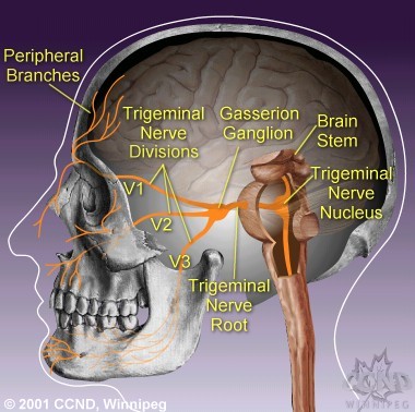
divisions which enervate the forehead and eye (ophthalmic V1), cheek (maxillary
V2) and lower face and jaw (mandibular V3). The trigeminal nerves function
in sensing facial touch, pain and temperature, as well as controlling muscles
used for chewing. The trigeminal nerve functions should be distinguished from
the facial nerve (cranial nerve VII), which controls all other facial movements.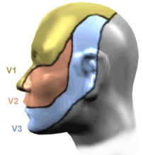
The
three divisions of the trigeminal nerve come together in an area called the
Gasserion ganglion. From there, the trigeminal nerve root continues
back towards the side of the brain stem, and inserts into the pons. Within
the brain stem, the signals traveling through the trigeminal nerve reach specialized
clusters of neurons called the trigeminal nerve nucleus. Information
brought to the brain stem by the trigeminal nerve is then processed before being
sent up to the brain and cerebral cortex, where a conscious perception of facial
sensation is generated.
Prepared by A.
M. Kaufmann & M.
Patel
© 2001 Centre for Cranial Nerve Disorders, Winnipeg, University of Manitoba,
Health Sciences Centre. The information provided on this web-site is intended
for educational purposes only, and should not be used to diagnose or treat a disease
or disorder. This information is not intended to substitute, supplement, or in
any way qualify the services or advice provided by a qualified health care professional.
Please consult with a certified health care professional before pursuing any form
of medical action. Duplication in any part or form of this document is strictly
prohibited. All rights reserved. For further information please read our disclaimer.
Web-Site related inquiries can be directed to the Information
Provider.